Reflectance confocal microscopy (RCM) is a non-invasive test that gives specialists a detailed view of skin cells, helping with diagnosis, treatment decisions, and monitoring.
Reflectance confocal microscopy (RCM), also called confocal microscopy, is a non-invasive imaging test that lets your skin specialist see your skin cells in very fine detail.
It looks at the top two layers of the skin to check for abnormal or cancerous cells. RCM can help:
Diagnose harmless (non-cancerous) skin spots
Decide which spots may need a biopsy
Guide treatment for melanoma
Monitor how well treatment is working
RCM is different from other skin imaging methods, such as dermoscopy (which uses a light and 10x magnification during a skin check). RCM is a more advanced test that shows much higher magnification, right down to individual skin cells.
Confocal microscopy is done with a small handheld microscope connected to a computer. It uses a gentle laser light to magnify your living skin cells by up to 1000 times.
This allows your dermatologist to see your skin cells in great detail—similar to looking at them under a lab microscope—without needing to cut out a piece of skin.
In some cases, confocal microscopy can reduce the need for a biopsy, but it doesn’t always replace one. Your doctor may still recommend a biopsy after the test.
Confocal microscopy is the most accurate non-invasive test available for diagnosing melanoma. Research shows it can correctly identify melanoma about 92% of the time.
It is also very good at diagnosing harmless (benign) spots on the skin, such as those caused by sun exposure or ageing. This means many people can avoid having unnecessary biopsies. In fact, studies show that confocal microscopy reduces unnecessary biopsies by about 43% compared with standard skin checks. Fewer biopsies mean less pain, less scarring, and less worry for patients, as well as lower costs for both patients and the health system.
When a biopsy is needed, confocal microscopy can guide doctors to sample the most suspicious part of the spot, making it more likely to get an accurate result.
Confocal microscopy can also be used before surgery to help map out the exact edges of a melanoma. This is especially useful for melanomas with unclear borders, or those on the face, so doctors can remove all the cancer while protecting as much healthy skin as possible.
In addition, confocal microscopy can be used to monitor non-surgical treatments (such as creams or radiotherapy). It helps doctors check how well treatment is working, confirm when the skin has fully responded, and pick up any signs of melanoma coming back at an early stage.
Confocal microscopy is a specialised area of dermatology. Only skin specialists with advanced training are qualified to use it. This includes at least six months of extra study and a deep understanding of how the skin works. This training means your doctor can use the test safely and give you accurate results.
Confocal microscopy is a safe and painless test that can be used on most people, including children and pregnant women. You don’t need to prepare for it, and it doesn’t leave any marks or scars.
Your dermatologist will carefully decide which spots on your skin may benefit from this test, usually during a skin check or dermoscopy. They will always explain why the test is recommended and what it can show.
Because the test only looks at the top layers of skin (about 0.2–0.3mm deep), it isn’t suitable for every type of lesion. It cannot be used for:
If you’ve recently had surgery or used a cream treatment for melanoma, your doctor may suggest waiting about three months before having confocal microscopy.
Confocal microscopy is especially helpful when there’s some uncertainty about a spot and your dermatologist needs more information. If the diagnosis is already clear, or if a biopsy is definitely needed, your doctor will explain why this test isn’t required and guide you through the next steps. You can feel confident that your care is always tailored to what is safest and most accurate for you.
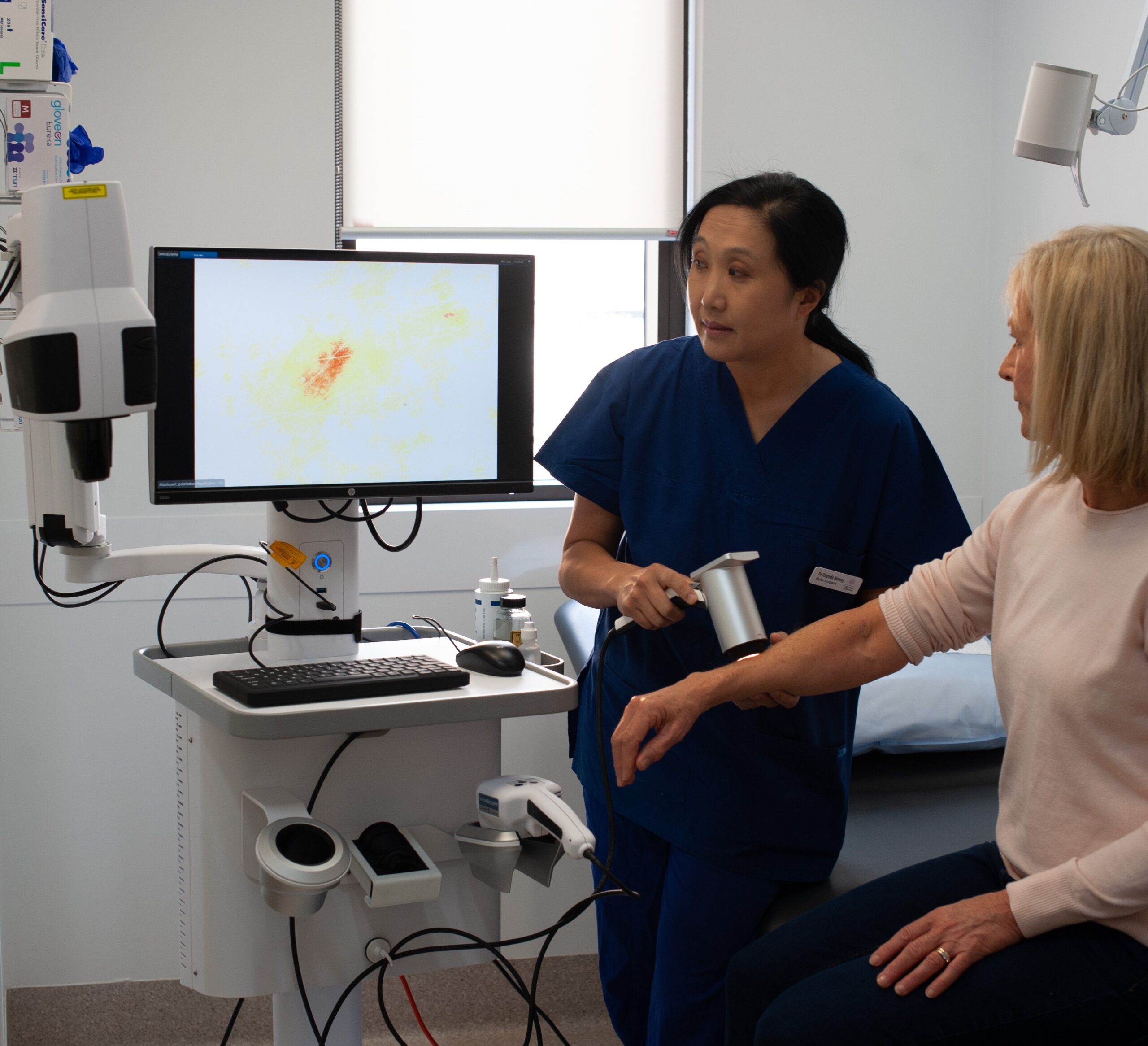
Confocal microscopy is a safe, painless, and non-invasive test. It usually takes about 15 to 30 minutes.
For the procedure, you’ll be asked to sit or lie still in a comfortable, darkened room while images of your skin are taken. The area of skin being checked will be gently cleaned. If needed, a small amount of hair may be removed, and a little gel applied to the skin.
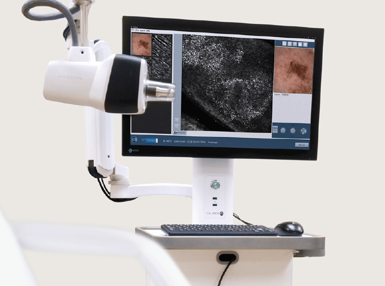
Depending on the type of microscope used, your dermatologist will either:
Most of the time, your dermatologist will be able to discuss the results with you straight away. Occasionally, the images may need to be reviewed in more detail, and your doctor will explain when and how you’ll receive the results.
You don’t need to do anything special to prepare, but it can help to wear loose, comfortable clothing so the area of skin can be easily accessed.
You can feel reassured that the procedure is simple, comfortable, and designed to give your doctor the clearest possible information about your skin without the need for surgery.
Confocal microscopy is a highly specialised test and is currently available only at a few major specialist centres in Australia.
Although the test can help reduce the need for unnecessary biopsies, it is not yet covered by Medicare. In some cases, it may be covered by the Department of Veterans’ Affairs or by certain private health insurers.
If you are recommended for this test, your dermatologist will explain the costs upfront and discuss any rebates or support options that may apply. This way, you’ll know exactly what to expect before proceeding.
Certain factors can make conducting and analysing confocal microscopy more difficult.